Introduction: Navigating the Rarity and Severity of Amniotic Embolism
Pregnancy and childbirth, while often joyous events, carry inherent risks. Among the most devastating and unpredictable complications is Amniotic Embolism (AE), also known as Anaphylactoid Syndrome of Pregnancy. This rare but catastrophic obstetric emergency can occur during labor, delivery, or immediately postpartum, striking suddenly and without warning. AE is characterized by a rapid onset of cardiopulmonary collapse and severe coagulopathy (blood clotting disorder) in the mother, triggered by the entry of amniotic fluid or fetal cells into the maternal bloodstream. Its rarity makes it less familiar to the general public, yet its profound severity and high mortality rate necessitate a thorough understanding among healthcare professionals and expectant families.
The term "embolism" in this context refers to the obstruction of blood vessels, but AE is far more complex than a simple blockage. It's now understood as an acute, severe anaphylactoid reaction, meaning the mother's body reacts as if to an allergen, leading to a cascade of inflammatory and clotting responses. This article delves into the critical aspects of Amniotic Embolism, focusing particularly on its survival rate, the factors influencing prognosis, and the comprehensive medical strategies employed to manage this life-threatening condition. We will explore the symptoms that signal its onset, the underlying mechanisms, diagnostic approaches, and the intensive treatments required to save both maternal and fetal lives. Understanding AE is crucial for improving outcomes and providing support to those affected by this medical enigma.
What is Amniotic Embolism (AE)?
Amniotic Embolism is a severe, life-threatening obstetric emergency that occurs when amniotic fluid, fetal cells, hair, or other debris from the fetus or placenta enters the mother's bloodstream. While the presence of amniotic fluid in maternal circulation is relatively common and often asymptomatic, in a small percentage of cases, it triggers an extreme, systemic inflammatory and anaphylactoid reaction. This reaction is not merely a mechanical obstruction but a complex immunological response that leads to a rapid and profound disruption of the mother's cardiovascular and respiratory systems, followed by a severe bleeding disorder.
The Pathophysiology of AE: A Two-Phase Event
The current understanding of AE describes a two-phase process:
- Phase 1: Cardiopulmonary Collapse. The initial phase is characterized by acute respiratory distress and cardiovascular collapse. The entry of amniotic fluid components into the maternal circulation causes a sudden increase in pulmonary artery pressure, leading to right ventricular failure. This is often accompanied by profound hypotension (low blood pressure), hypoxemia (low blood oxygen), and cardiac arrest. The exact mechanism is thought to involve vasoactive substances within the amniotic fluid or released by the maternal immune system in response to the fluid, causing pulmonary vasoconstriction and myocardial dysfunction.
- Phase 2: Hemorrhage and Coagulopathy. If the mother survives the initial cardiopulmonary collapse, the second phase typically involves severe coagulopathy, specifically disseminated intravascular coagulation (DIC). This is a complex disorder where the body's clotting system becomes overactivated, leading to widespread formation of tiny blood clots throughout the body. Paradoxically, this widespread clotting consumes clotting factors and platelets at an alarming rate, leading to a severe inability to form clots where needed, resulting in massive hemorrhage, often from the uterus, surgical sites, or intravenous access points. This uncontrolled bleeding significantly exacerbates the life-threatening nature of AE.
AE is a diagnosis of exclusion, meaning it's often diagnosed after other more common causes of maternal collapse or hemorrhage have been ruled out. Its unpredictable nature and rapid progression make it one of the most feared complications in obstetrics.
Symptoms of Amniotic Embolism
The onset of Amniotic Embolism is typically sudden and dramatic, often occurring without any prior warning signs. Symptoms can vary slightly but generally follow a rapid progression of severe systemic distress. Recognizing these symptoms immediately is crucial for prompt intervention.
Key Symptoms Include:
- Sudden Respiratory Distress: This is often one of the first and most striking symptoms. The mother may experience sudden shortness of breath (dyspnea), rapid breathing (tachypnea), and a feeling of impending doom. Oxygen saturation levels drop precipitously.
- Acute Hypotension/Cardiovascular Collapse: A rapid and severe drop in blood pressure is characteristic. This can quickly progress to profound shock and cardiac arrest. The heart rate may initially increase (tachycardia) as the body tries to compensate, but can become erratic (arrhythmias) or cease.
- Cyanosis: A bluish discoloration of the skin, lips, and nail beds due to lack of oxygen in the blood.
- Altered Mental Status: Confusion, disorientation, loss of consciousness, or seizures can occur due to lack of oxygen to the brain.
- Fetal Distress: As the mother's oxygen supply and cardiovascular function falter, the fetus is also profoundly affected. Signs of fetal distress, such as sudden and severe bradycardia (slow heart rate) or loss of fetal heart rate variability, are common and often necessitate immediate emergency delivery.
- Coagulopathy and Hemorrhage: If the initial cardiopulmonary phase is survived, profound bleeding typically ensues. This can manifest as excessive bleeding from the vagina, the site of a C-section incision, intravenous lines, or even from mucous membranes. This uncontrolled bleeding is due to disseminated intravascular coagulation (DIC), where the body's clotting factors are rapidly consumed.
- Uterine Atony: The uterus may fail to contract adequately after delivery, contributing to severe postpartum hemorrhage.
- Chills and Rigors: Although less common, some women may experience these symptoms.
The rapid progression of these symptoms, often within minutes, underscores the urgency required for diagnosis and treatment. The multidisciplinary team involved in an obstetric setting must be acutely aware of these signs to initiate life-saving measures without delay.
Causes and Risk Factors of Amniotic Embolism
Despite extensive research, the exact cause of Amniotic Embolism remains largely unknown, and it is largely considered an unpredictable event. It is not caused by anything the mother or healthcare provider does or fails to do. The prevailing theory, as mentioned, is an acute anaphylactoid reaction to fetal material entering the maternal circulation, rather than a simple mechanical obstruction.
While the direct cause is elusive, several factors have been identified that may increase the risk, though it's important to remember that AE can occur in women with no identifiable risk factors:
- Advanced Maternal Age: Women over 35 years old appear to have a slightly higher risk.
- Multiparity: Having had multiple previous pregnancies.
- Rapid or Forceful Labor: Very fast labor or the use of uterotonic agents (medications to strengthen contractions) might increase the likelihood of amniotic fluid entering the maternal circulation.
- Fetal Distress: Conditions that cause stress to the fetus, such as meconium-stained amniotic fluid, may be associated.
- Placental Abnormalities: Conditions like placenta previa (placenta covering the cervix) or placental abruption (premature separation of the placenta from the uterus) are sometimes linked.
- Preeclampsia/Eclampsia: These hypertensive disorders of pregnancy may be associated with an increased risk.
- Polyhydramnios: Excessive amniotic fluid.
- Cervical Lacerations/Uterine Trauma: Any breach in the integrity of the utero-placental barrier could theoretically facilitate the entry of amniotic fluid.
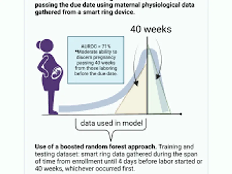
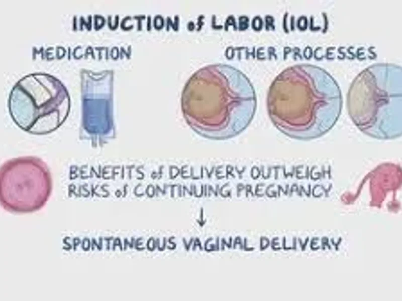
- Induced Labor: Some studies suggest a possible association, though this remains debated.
- Caesarean Section or Operative Vaginal Delivery: Surgical procedures or interventions during delivery may increase the chance of amniotic fluid entering the bloodstream, particularly if uterine veins are exposed.
- Medical Induction of Labor: While not a direct cause, the process of inducing labor might increase the risk of situations where AE could occur.
- Maternal Medical Conditions: Some chronic conditions might marginally increase susceptibility, but this is not well-established.
It is crucial to emphasize that AE is extremely rare, occurring in approximately 1 in 40,000 to 1 in 80,000 deliveries. Even with the presence of multiple risk factors, the vast majority of women will not experience AE. These risk factors are associations, not direct causes, and their presence does not mean AE will occur. The focus remains on rapid recognition and management rather than prevention based on risk factor modification, as AE is largely unpreventable.
Diagnosis of Amniotic Embolism
Diagnosing Amniotic Embolism is challenging because there is no specific diagnostic test. It is primarily a clinical diagnosis, meaning it's made based on the sudden onset of characteristic symptoms and the exclusion of other potential causes of maternal collapse or hemorrhage. The rapid progression of AE often means that definitive diagnosis occurs retrospectively, after the immediate crisis has been managed or, sadly, after maternal death.
Key Aspects of Clinical Diagnosis:
- Sudden Onset: The hallmark is the abrupt and unexpected development of symptoms during labor, delivery, or within 30 minutes postpartum.
- Triad of Symptoms: The classic presentation involves the rapid onset of:
- Hypotension/Cardiopulmonary Arrest: Severe drop in blood pressure leading to shock or cardiac arrest.
- Hypoxemia/Respiratory Distress: Difficulty breathing, low oxygen levels, and often cyanosis.
- Coagulopathy/Hemorrhage: Unexplained, severe bleeding due to a clotting disorder.
- Exclusion of Other Causes: It's essential to rule out other conditions that can present similarly, such as:
- Anaphylaxis (allergic reaction)
- Sepsis (severe infection)
- Pulmonary embolism (blood clot in the lung, often from deep vein thrombosis)
- Eclampsia (severe preeclampsia leading to seizures)
- Local anesthetic systemic toxicity
- Cardiac arrest from other causes (e.g., myocardial infarction)
- Severe postpartum hemorrhage from uterine atony or trauma
Supportive Diagnostic Clues (often retrospective):
While not for immediate diagnosis, certain findings can support a diagnosis of AE:
- Laboratory Tests: Blood tests will show evidence of severe coagulopathy:
- Reduced platelet count (thrombocytopenia)
- Prolonged prothrombin time (PT) and activated partial thromboplastin time (aPTT)
- Decreased fibrinogen levels
- Elevated D-dimer (indicating widespread clot formation and breakdown)
- Fetal Squames in Maternal Blood: Historically, the presence of fetal squamous cells (skin cells) in maternal pulmonary capillaries or central venous blood samples was considered diagnostic. However, this finding is not universally present, can be found in asymptomatic women, and is usually only detectable post-mortem or retrospectively. It is no longer considered a definitive diagnostic criterion for acute management.
- Echocardiography: May show right ventricular dysfunction and pulmonary hypertension.
Given the urgency, the focus is on rapid clinical assessment and initiating empirical treatment for AE based on the characteristic presentation while simultaneously attempting to rule out other conditions. A high index of suspicion in any case of sudden maternal collapse during the peripartum period is paramount.
Treatment Options for Amniotic Embolism
The management of Amniotic Embolism is one of the most critical and complex challenges in obstetric medicine, requiring immediate, aggressive, and multidisciplinary intervention. Treatment is primarily supportive, aimed at maintaining maternal life and managing the catastrophic physiological changes that occur.
Immediate Life-Saving Measures (The "AET Kit" or "Code AE"):
Many institutions now have a pre-prepared "Amniotic Embolism Treatment Kit" or a designated "Code AE" protocol to streamline the response.
- Activation of Emergency Response: Immediately call for help – activate the hospital's obstetric emergency team, anesthesiology, critical care, hematology, and blood bank.
- Cardiopulmonary Resuscitation (CPR): If cardiac arrest occurs, initiate high-quality CPR immediately. Maternal CPR differs slightly, requiring uterine displacement to the left to relieve pressure on the vena cava.
- Respiratory Support: Secure the airway and provide 100% oxygen. Intubation and mechanical ventilation are almost always necessary due to severe hypoxemia and respiratory failure.
- Cardiovascular Support: Administer intravenous fluids cautiously to avoid fluid overload, which can worsen pulmonary edema. Vasopressors (e.g., norepinephrine, epinephrine) are crucial to combat severe hypotension and maintain blood pressure. Inotropic agents may be used to improve cardiac contractility.
- Correction of Coagulopathy and Hemorrhage Management: This is a cornerstone of AE management. Massive transfusion protocols should be activated immediately.
- Blood Products: Aggressive transfusion of packed red blood cells (PRBCs) to address blood loss and improve oxygen-carrying capacity.
- Fresh Frozen Plasma (FFP): To replace clotting factors.
- Platelets: To correct thrombocytopenia.
- Cryoprecipitate: To replace fibrinogen and factor VIII.
- Tranexamic Acid (TXA): An antifibrinolytic agent to help stabilize clots and reduce bleeding.
- Emergency Delivery: If the fetus is still in utero, an immediate emergency C-section (perimortem C-section if cardiac arrest) is often necessary. This serves two purposes: to save the baby from severe fetal distress due to maternal hypoxemia and hypotension, and to potentially improve maternal resuscitation by relieving pressure on the maternal great vessels and diaphragm, thereby improving cardiac output.
- Uterine Atony Management: Postpartum hemorrhage due to uterine atony is common. Uterotonic agents (e.g., oxytocin, methylergonovine, carboprost) should be administered, and if these fail, surgical interventions like uterine tamponade, B-Lynch suture, or even hysterectomy may be required as a last resort to control bleeding.
- Monitoring and Intensive Care: Continuous, aggressive monitoring in an Intensive Care Unit (ICU) is essential. This includes invasive hemodynamic monitoring (arterial line, central venous pressure, possibly pulmonary artery catheter), continuous cardiac monitoring, pulse oximetry, and frequent blood gas analysis and coagulation studies.
- Adjunctive Therapies: Some institutions may use specific agents, though evidence for their routine use is limited:
- Atropine, Ondansetron, Ketorolac (AOK Protocol): Some anecdotal reports suggest this combination may help modulate the inflammatory response, but it is not universally adopted.
- Corticosteroids: May be considered to dampen the inflammatory response.
- Nitric Oxide or Prostacyclin Analogs: For severe pulmonary hypertension.
- Emotional and Psychological Support: For survivors and their families, the experience is traumatic. Long-term psychological support, including counseling for PTSD, is often necessary.
The rapid mobilization of a highly skilled multidisciplinary team is the single most important factor in successful management. Every minute counts, and a well-rehearsed protocol can significantly improve the chances of survival.
Amniotic Embolism Survival Rate and Prognosis
Amniotic Embolism has historically been associated with an extremely high mortality rate, making it one of the leading direct causes of maternal death. However, advancements in critical care, rapid response protocols, and a deeper understanding of its pathophysiology have led to improvements in survival rates over recent decades.
Historical vs. Modern Survival Rates:
- Historically: The mortality rate for AE was reported to be as high as 60-80% or even higher, often cited as one of the most lethal obstetric complications.
- Modern Era: While still very high, more recent studies and large population registries report maternal mortality rates ranging from 13% to 26%. This significant improvement is attributed to faster recognition, immediate access to advanced critical care, improved resuscitation techniques, and aggressive management of coagulopathy. It's important to note that even with these improvements, AE remains a condition with a substantial risk of death or severe morbidity.
Factors Influencing Survival and Prognosis:
- Speed of Diagnosis and Intervention: The most critical factor. Rapid recognition of symptoms and immediate initiation of resuscitation and supportive measures are paramount. Delays significantly worsen outcomes.
- Availability of Resources: Access to a fully equipped intensive care unit (ICU), a well-stocked blood bank, and a multidisciplinary team (obstetricians, anesthesiologists, critical care specialists, hematologists) is crucial. Hospitals with high levels of obstetric care tend to have better outcomes.
- Severity of Initial Presentation: Patients who experience immediate cardiac arrest have a lower survival rate compared to those who primarily present with respiratory distress and hypotension.
- Extent of Coagulopathy: The severity and responsiveness of the disseminated intravascular coagulation (DIC) to blood product transfusion also play a significant role. Uncontrolled hemorrhage is a major cause of death.
- Fetal Outcome: Fetal survival is highly dependent on maternal survival and the speed of delivery once AE occurs. If the mother experiences cardiac arrest, perimortem C-section within 5 minutes is critical for fetal survival and can also aid maternal resuscitation.
Long-Term Prognosis for Survivors:
Even for those who survive the acute phase of AE, the long-term prognosis can be challenging. Many survivors face significant morbidity due to the severity of the initial event and the extensive resuscitation required.
- Neurological Impairment: Hypoxic brain injury due to cardiac arrest or prolonged hypotension is a major concern. Survivors may experience a range of neurological deficits, from mild cognitive impairment to severe anoxic brain injury, requiring long-term rehabilitation.
- Multi-Organ Dysfunction: Kidneys, liver, and lungs can be affected, potentially leading to chronic organ damage or dysfunction.
- Psychological Trauma: Survivors and their partners often experience severe psychological distress, including post-traumatic stress disorder (PTSD), anxiety, and depression. The suddenness and life-threatening nature of AE, coupled with potential neurological deficits or the loss of their baby, can have profound mental health impacts.
- Future Pregnancies: While a subsequent AE is extremely rare, women who have survived AE require careful counseling regarding future pregnancies. The decision to have another child involves careful consideration of the psychological impact and potential, albeit very low, recurrence risk.
The journey for an AE survivor and their family is often long and arduous, requiring extensive medical, rehabilitative, and psychological support. Support groups and specialized counseling can be invaluable resources.
Long-term Effects for Survivors
Surviving an amniotic embolism is a testament to extraordinary medical care and the resilience of the human body, but it is rarely without significant long-term consequences. The profound physiological insult to the mother's body can lead to a range of chronic health issues, both physical and psychological.
Physical Long-term Effects:
- Neurological Impairment: This is one of the most devastating potential long-term effects. The brain is highly sensitive to oxygen deprivation. Even brief periods of cardiac arrest or severe hypotension can lead to hypoxic-ischemic encephalopathy. This can manifest as:
- Cognitive Deficits: Problems with memory, concentration, executive function, and learning.
- Motor Impairment: Weakness, spasticity, coordination problems, or even paralysis, requiring extensive physiotherapy.
- Seizure Disorders: New-onset epilepsy can develop after brain injury.
- Speech and Swallowing Difficulties: Dysarthria or dysphagia, requiring speech therapy.
- Cardiopulmonary Issues: While the acute phase involves severe cardiopulmonary compromise, some survivors may experience long-term cardiac dysfunction or pulmonary issues. This could include persistent arrhythmias, decreased cardiac function, or chronic lung problems, especially if prolonged ventilation was required or if there was significant acute lung injury.
- Renal Dysfunction: Acute kidney injury is common during the critical phase of AE due to shock and hypoperfusion. While many recover, some may develop chronic kidney disease, requiring ongoing monitoring or, in severe cases, dialysis.
- Endocrine Dysfunction: Damage to the pituitary gland (Sheehan's syndrome) due to severe blood loss and shock can lead to long-term hormonal deficiencies, affecting thyroid function, adrenal function, and reproductive hormones.
- Uterine and Reproductive Health: If a hysterectomy was performed to control hemorrhage, this obviously impacts future fertility. Even without hysterectomy, the trauma to the uterus and surrounding tissues can lead to complications in future pregnancies, though recurrence of AE is exceedingly rare.
- Chronic Pain and Fatigue: The extensive medical interventions, surgeries, and prolonged recovery can lead to chronic pain, muscle weakness, and debilitating fatigue.
Psychological and Emotional Long-term Effects:
- Post-Traumatic Stress Disorder (PTSD): The sudden, life-threatening nature of AE, often coupled with the trauma of an emergency delivery, potential loss of the baby, and intensive care experience, makes PTSD a very common and significant long-term issue for survivors and their partners. Symptoms can include flashbacks, nightmares, avoidance behaviors, hypervigilance, and emotional numbness.
- Anxiety and Depression: These are prevalent, stemming from the trauma, the ongoing physical challenges, the impact on family life, and grief, especially if the baby did not survive.
- Grief and Loss: Survivors may grieve the loss of their health, their ability to have more children, the traumatic birth experience, or the loss of their baby. This grief can be complex and prolonged.
- Impact on Relationships: The emotional and physical toll can strain relationships with partners, family, and friends.
- Body Image Issues: Related to surgical scars (C-section, hysterectomy), weight changes, or other physical alterations.
Comprehensive follow-up care for AE survivors must be multidisciplinary, encompassing not only medical specialists (neurologists, nephrologists, cardiologists, endocrinologists) but also extensive psychological support, physical therapy, occupational therapy, and social work services. Support groups for AE survivors and their families can provide invaluable peer support and a sense of community.
Prevention of Amniotic Embolism
Unfortunately, Amniotic Embolism is largely considered an unpreventable event. Its unpredictable nature and the fact that it can occur in women with no identifiable risk factors mean that there are no definitive strategies to prevent its occurrence. Unlike many other obstetric complications, AE is not caused by medical error or negligence, nor is it typically influenced by lifestyle choices.
Why Prevention is Challenging:
- Unknown Etiology: The exact trigger for the severe anaphylactoid reaction remains elusive. While the entry of amniotic fluid into maternal circulation is a prerequisite, why it causes a catastrophic reaction in some and is asymptomatic in most others is not fully understood.
- Lack of Predictive Markers: There are no reliable screening tests or biomarkers that can predict which women are at risk of developing AE.
- Rarity: Its extreme rarity makes large-scale prevention studies difficult.
Focus on Preparedness and Management:
Given the unpreventable nature of AE, the focus in modern obstetrics shifts entirely to preparedness and rapid, effective management. The goal is to minimize morbidity and mortality once the event occurs.
- Education and Training: Healthcare providers, especially those in labor and delivery, need to be highly educated about the signs and symptoms of AE and trained in rapid response protocols. Regular drills and simulations (e.g., Code AE drills) are crucial for ensuring a coordinated and efficient team response.
- Multidisciplinary Team Readiness: Ensuring that a multidisciplinary team (obstetricians, anesthesiologists, critical care specialists, hematologists, blood bank personnel) can be rapidly mobilized is essential.
- Availability of Resources: Hospitals must have immediate access to a well-stocked blood bank (including all necessary blood products like PRBCs, FFP, platelets, cryoprecipitate), emergency medications (vasopressors, uterotonics), and advanced life support equipment.
- Established Protocols: Clear, concise, and easily accessible protocols for managing AE, including massive transfusion protocols, should be in place and regularly reviewed.
- Risk Factor Awareness (with caveats): While risk factors don't cause AE, being aware of them (e.g., advanced maternal age, C-section) might prompt a slightly higher index of suspicion in certain cases, though this should not detract from vigilance for all patients.
In essence, while we cannot prevent Amniotic Embolism from happening, we can significantly improve the chances of survival and reduce the severity of long-term complications through meticulous preparation, prompt recognition, and aggressive, standardized management. The best "prevention" in this context is readiness.
When to See a Doctor
Amniotic Embolism is an acute, emergency event that occurs during labor, delivery, or immediately postpartum. Therefore, it is not a condition where a pregnant woman would "see a doctor" in advance for symptoms of AE. Instead, it is a sudden medical catastrophe that demands immediate, in-hospital emergency medical intervention.
What Expectant Mothers and Support Persons Should Know:
While you cannot prevent AE or recognize its subtle onset before it becomes critical, it is important for expectant mothers and their support persons to be aware of the following:
- During Labor and Delivery: If you or your support person notice any sudden, severe, and unexplained changes in your condition during labor or immediately after birth, such as:
- Sudden, extreme shortness of breath or difficulty breathing
- Feeling dizzy, lightheaded, or like you might pass out
- Sudden, severe chest pain or discomfort
- Sudden confusion or disorientation
- Unexplained heavy bleeding after delivery that seems excessive or unusual
It is crucial to alert your healthcare team immediately. While these symptoms could be indicative of many less severe conditions, in the context of AE, every second counts. Your medical team is trained to recognize and respond to such emergencies.
- Trust Your Instincts: If you feel that something is profoundly wrong, do not hesitate to voice your concerns to your nurses or doctors.
What Healthcare Providers Should Do:
For healthcare providers, the imperative is to maintain a high index of suspicion for AE in any peripartum woman presenting with sudden, unexplained cardiopulmonary collapse, severe hypoxemia, or uncontrollable hemorrhage. Immediate actions include:
- Activate Emergency Protocols: Call for a rapid response team, Code Blue, or specific AE protocol immediately.
- Initiate Resuscitation: Begin CPR if necessary, provide oxygen, establish IV access, and initiate fluid and vasopressor support.
- Manage Airway and Breathing: Prepare for intubation and mechanical ventilation.
- Address Hemorrhage and Coagulopathy: Activate massive transfusion protocol and administer blood products.
- Consider Emergency Delivery: If the fetus is still in utero, prepare for immediate C-section.
In summary, Amniotic Embolism is an emergency that necessitates immediate action by a trained medical team within a hospital setting. There is no pre-hospital management or